Blue Nevus Skin Cancer Clinic Brisbane
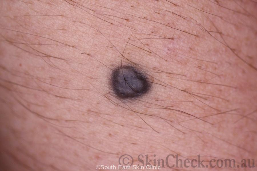
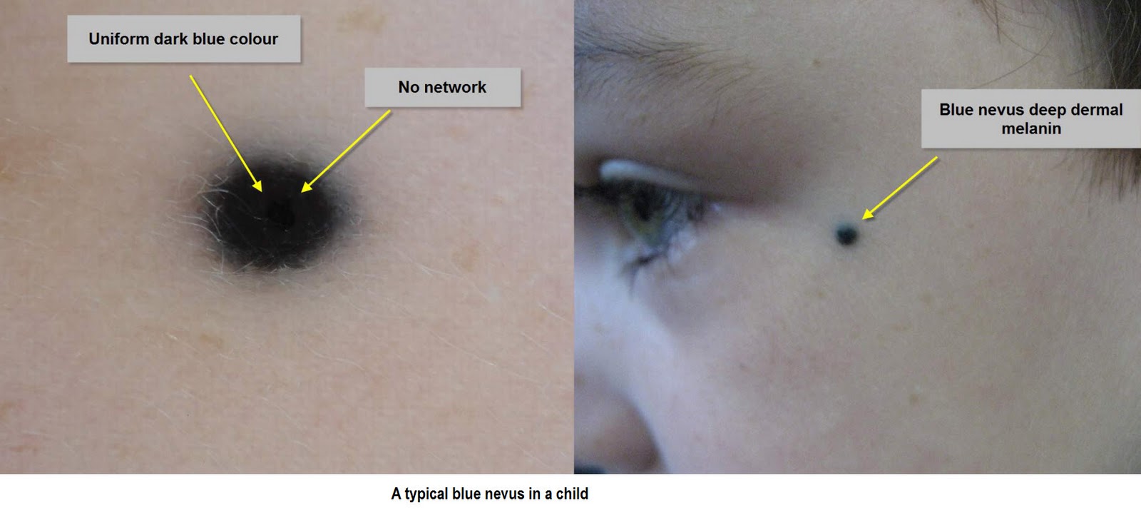
A blue naevus is a common type of melanocytic naevus in which the pigment is situated deep in the dermis. It is also known as a blue neuronaevus and a dermal melanocytoma. What are the clinical features of a blue naevus? A blue naevus is a well- circumscribed round or oval macule, papule or nodule of a uniform steel blue colour.

Naevus coeruleus (blue nevus)
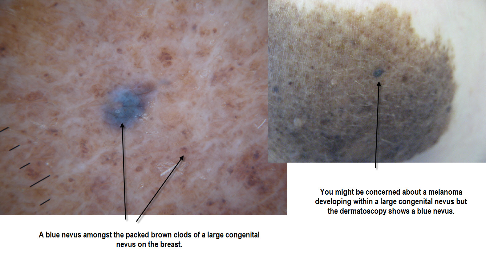
Blue nevus is a benign melanocytic lesion whose most frequent variants are dendritic (common) blue nevus and cellular blue nevus. Atypical cellular blue nevus presents an intermediate histopathology between the typical and a rare variant of malignant blue nevus/melanoma arising in a cellular blue nevus. An 8-year-old child presented a pigmented.
Melanocytic Nevi Blue Nevus Overview Perri Dermatology
Browse 136 blue nevus photos and images available, or start a new search to explore more photos and images. 3 NEXT Browse Getty Images' premium collection of high-quality, authentic Blue Nevus stock photos, royalty-free images, and pictures. Blue Nevus stock photos are available in a variety of sizes and formats to fit your needs.

Pathology Outlines Common blue nevus
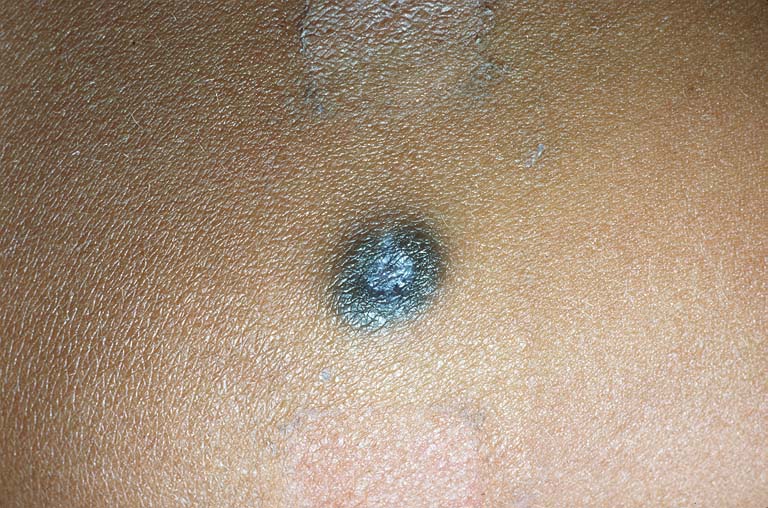
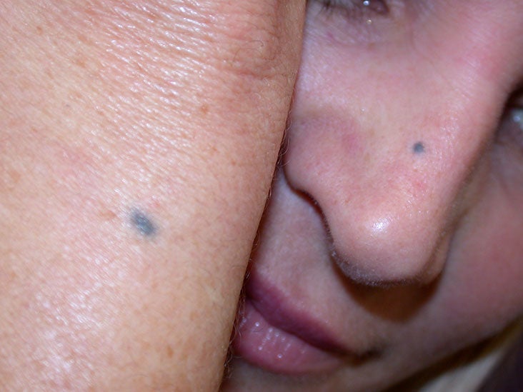
The blue nevus is a solitary blue-colored mole that can present at birth or develop later on in life. It can remain unchanged throughout the duration of a person's life. The blue color, or ceruloderma, is caused by the Tyndall effect when light is preferentially scattering shorter wavelengths by melanin found in the dermis of the skin.

Blue Nevus Stock Image C043/0668 Science Photo Library
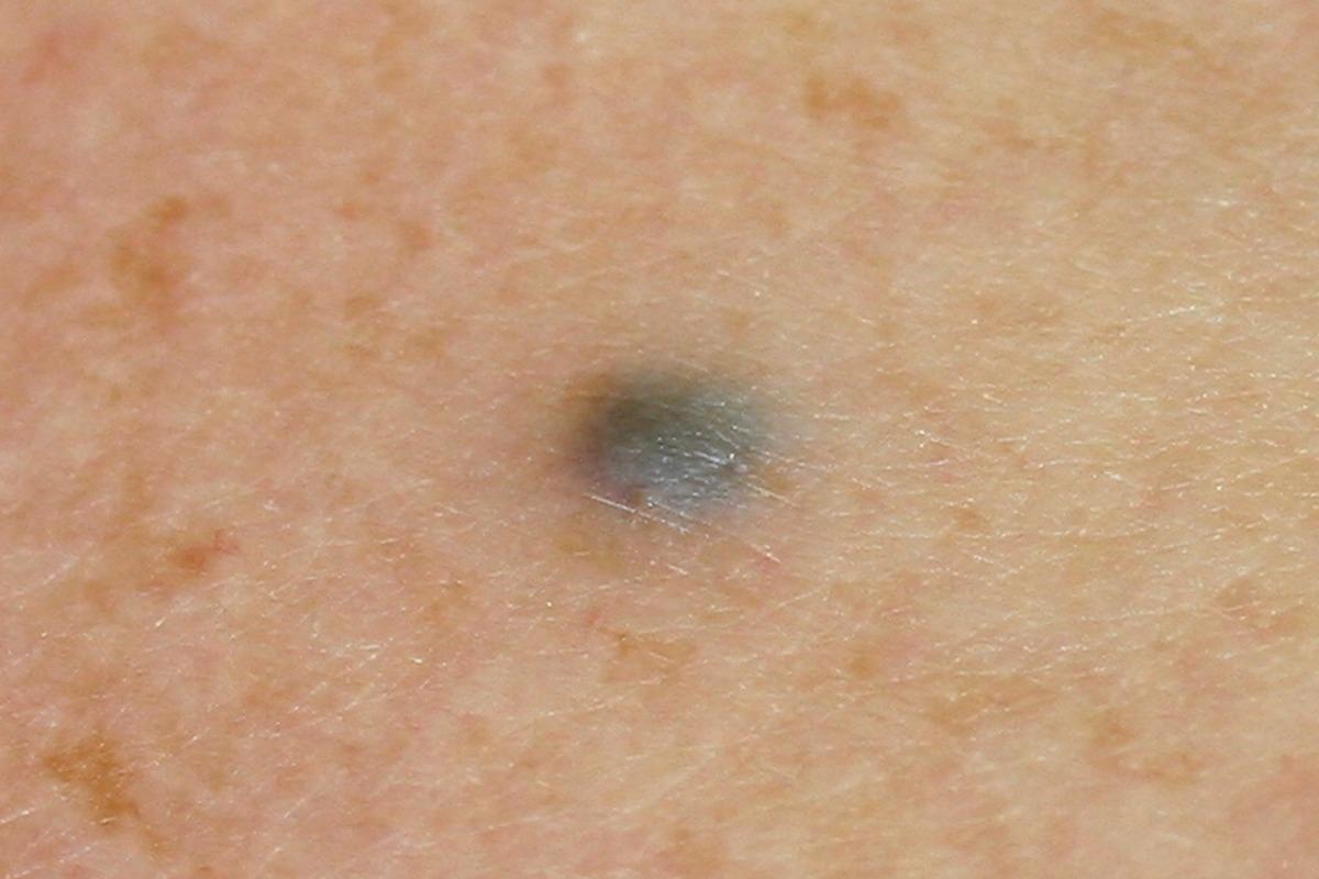
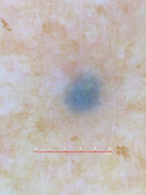
A blue nevus is a type of coloured mole, typically a single well-defined blue-black bump. [1] [2] The blue colour is caused by the pigment being deep in the skin. [4] Diagnosis is by visualisation and dermoscopy. [4] A biopsy is sometimes performed, or the whole lesion surgically removed. [3]
blue nevi pictures pictures, photos
Microscopic images of blue nevi. The epidermis is uninvolved in common blue nevi; however, peri-appendageal growth is not uncommon. The common blue nevus is composed of a variably dense proliferation of spindle-shaped and dendritic melanocytes with an associated infiltrate of pigment-laden macrophages ("melanophages") (Figure 1, B).

Blue nevus Stock Image C051/6921 Science Photo Library
A blue nevus is a blue-colored mole that can be congenital or acquired. A common blue nevus may appear flat or dome-shaped with a color ranging from blue-grey to blue-black.
What is Blue Rubber Bleb Nevus Syndrome? Family Medicine News
Conclusions. Blue nevus is a benign neoplasm composed of aberrant melanocytes located in the dermis, which can be found in 0.5%-4% of the white population. It is usually acquired during the second decade of life, presenting as a macule, papule, nodule, or plaque on the buttocks, face, scalp, hands, or feet.

Blue Nevus Stock Image C022/2095 Science Photo Library
The term "blue nevus" describes a group of skin lesions characterized by dermal proliferation of melanocytes presenting as blue to black nodules on the head, extremities, or buttocks. In most cases, they are acquired and present as a solitary lesion but may also be congenital and appear at multiple sites.

Blue Nevus Academic Dermatology of Nevada
Videos Figures Images Quizzes Symptoms View All Resources. RESOURCES 3D Models Abbreviations Lab Tests Audio Case. This photo shows a blue nevus composed of dark pigmented cells (melanocytes). DermPics/SCIENCE PHOTO LIBRARY.

Dermoscopy of Melanocytic Neoplasms Blue Nevi Dermatology JAMA Dermatology JAMA Network
Pictures Symptoms A common blue nevus presents as a smooth oval or circular mole. A blue nevus is typically blue or gray, but it can sometimes be yellowish-brown. The main.
Blue nevus on the left forearm. (A) Enlarged “tape de Openi
Blue nevus. This is a dark blue-colored mole that can appear anywhere on the skin and doesn't change with age. They are usually present at birth but can develop later in life. Image Source: © Richard Usatine, MD Text Source: American Osteopathic College of Dermatology Dermatology Advisor

Clinical and Dermoscopic Features of Agminated Blue Nevus Dermatology JAMA Dermatology
Blue nevus is characterized by a dermal proliferation of melanocytes Common (dendritic) blue nevus: Dermal proliferation of melanocytes (dermal melanocytosis) presenting as a blue to black nodule; sometimes multiple; amelanotic lesions may occur ( StatPearls: Blue Nevus [Accessed 30 August 2021] ) Mostly acquired but it can rarely be congenital

Naevus coeruleus (blue nevus)
File:Blue nevus fig 1a.jpg Clinical characteristics BN are benign dermal melanocytic proliferation arising from neural-crest melanocytes that likely became arrested in migration during embryogenesis. Their prevalence ranges between 1-2% in Caucasians and 3-5% in Asians.
Blue nevus Pictures, diagnosis, removal, and more
Browse 1,100+ blue nevus stock photos and images available, or start a new search to explore more stock photos and images. Sort by: Most popular Young Woman in Winter A young, multi-ethnic woman outdoors in winter. Image of baby Mongolian spot Dark colour Blue neuronevus mole aka dermal melanocytoma, a type.
Dermoscopy Made Simple Blue nevus
Nov. 17, 2022 What Is a Nevus? A nevus is a common, colored growth on or in your eye. Sometimes called a freckle of the eye, it is similar to a mole on your skin. A nevus (plural: nevi) can be in the front of your eye, around the iris, or under the retina at the back of the eye. What Causes Nevi? A nevus is made up of cells called melanocytes.