Inguinal Canal Anatomy
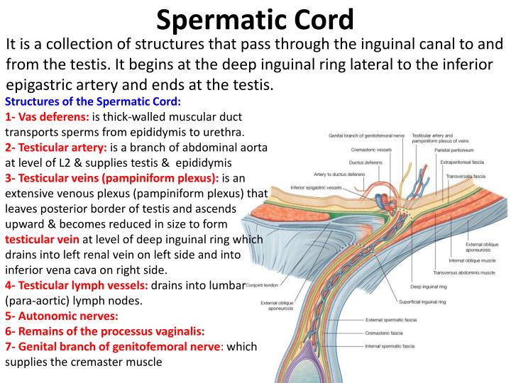
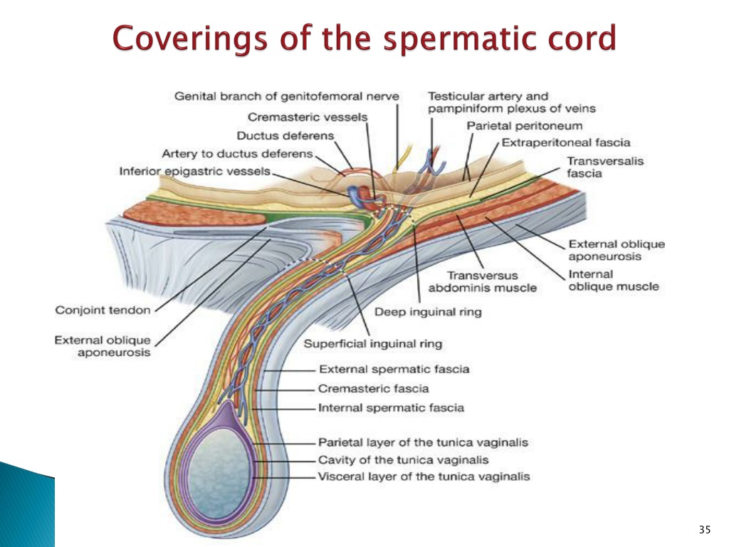
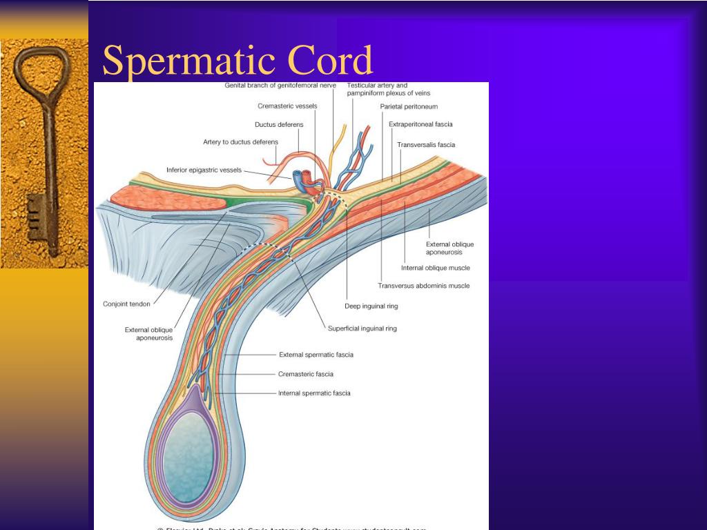
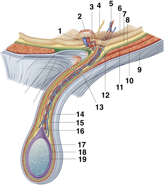
Contents of the spermatic cord include the ductus deferens, artery of the ductus deferens, testicular artery, cremasteric artery, pampiniform plexus of veins, genital branch of the genitofemoral nerve, lymphatics and sympathetic and parasympathetic nerve fibers.

1. Anatomy of the Man SimpleMed Learning Medicine, Simplified
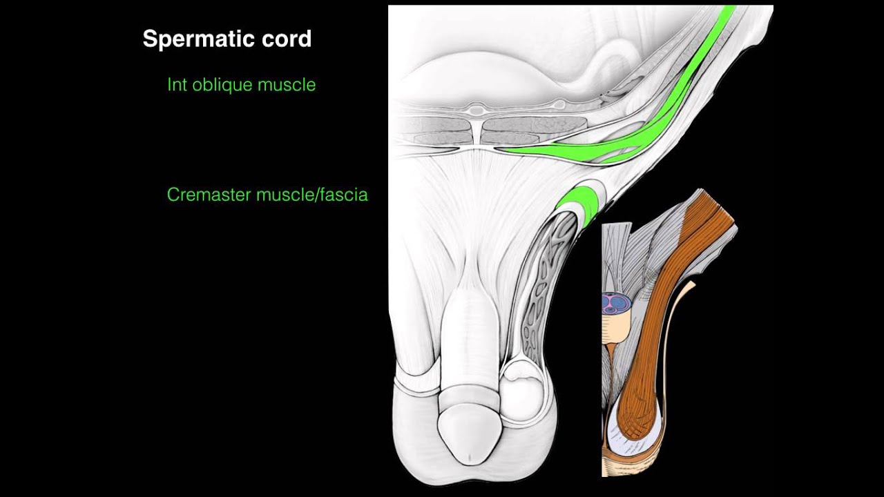
The spermatic cord ( TA : funiculus spermaticus) is the tubular structure that suspends the testes and epididymis in the scrotum from the abdominal cavity. Gross anatomy Course The spermatic cord arises at the deep inguinal ring, passes through the inguinal canal and exits at the superficial inguinal ring into the scrotum 3.
Contents Of Spermatic Cord / abdomen Biology 4930 with Dr. E at State The
Introduction The inguinal canal is a passage in the anterior abdominal wall that conveys several structures including nerves (ilioinguinal, genital branch of the genitofemoral nerve), spermatic cord (males) and round ligament (females). There is an inguinal canal present on both sides of the abdomen, running parallel to the inguinal ligament.
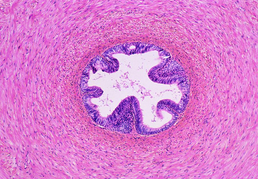
Light Micrograph Of A Section Of Spermatic Cord. Photograph by Astrid & Hannsfrieder Michler
Handy mnemonics to recall the contents of the spermatic cord are: Papers Don't Contribute To A Good Specialist Level 3 arteries, 3 nerves, 3 fascias, 3 other things Mnemonics Papers Don't Contribute To A Good Specialist Level P: pampiniform plexus D: ductus deferens C: cremasteric artery T: testicular artery
Contents of the scrotum Epididymis, vas deferens, spermatic cord and cremaster muscle
Contents of spermatic cord is a topic frequently asked in medical exams. The most common question on this topic is something like "all of the following are contents of spermatic cord except". In this post, we will memorize the contents of spermatic cord using a simple mnemonic. Remember : Plenty of Dick Contributes To Good Sex Life

1942 Inguinal Canal and Coats of Spermatic Cord Original Vintage Print Anatomy Medical Decor
One of the most important diagnostic tools is the spermatic cord block, used to localize the source of the pain to the scrotal contents (the distribution of pain fibers from the spermatic cord). A spermatic cord block is indicated in the absence of an obvious alternative source for the pain. Local anesthetic is administered into the spermatic.

spermatic cord anatomy 3d anatomy of spermatic cord contents covering of spermatic cord
Contents Anatomy Landmarks Borders Contents Inguinal canal in females Inguinal canal in males Embryology Clinical application: Inguinal hernia Types of inguinal hernias Symptoms of an inguinal hernia Examination of a hernia Sources + Show all Anatomy Landmarks
PPT The abdominal wall and inguinal region PowerPoint Presentation, free download ID9468778
Pathologic conditions that affect the spermatic cord (SC) are relatively common in everyday clinical practice. These conditions range from asymptomatic abnormalities that are incidentally discovered to common causes of emergency medicine visits. Visits for male genitourinary complaints comprise 0.5%-2.5% of all emergency medicine visits ( 1, 2 ).

Instant Anatomy Abdomen Inguinal region Spermatic cord & contents of
The Spermatic Cord (funiculus spermaticus) extends from the abdominal inguinal ring, where the structures of which it is composed converge, to the back part of the testis.In the abdominal wall the cord passes obliquely along the inguinal canal, lying at first beneath the Obliquus internus, and upon the fascia transversalis; but nearer the pubis, it rests upon the inguinal and lacunar ligaments.

GROSS II, EXAM III, Fig. 4.47 spermatic cord Diagram Quizlet
The spermatic cord refers to a collection of vessels, nerves and ducts that run to and from the testes. They are surrounded by fascia, forming a cord-like structure. This article will look at the anatomy of the spermatic cord - its anatomical course, contents, and clinical correlations. Anatomical Course
PPT Thorax PowerPoint Presentation, free download ID963761
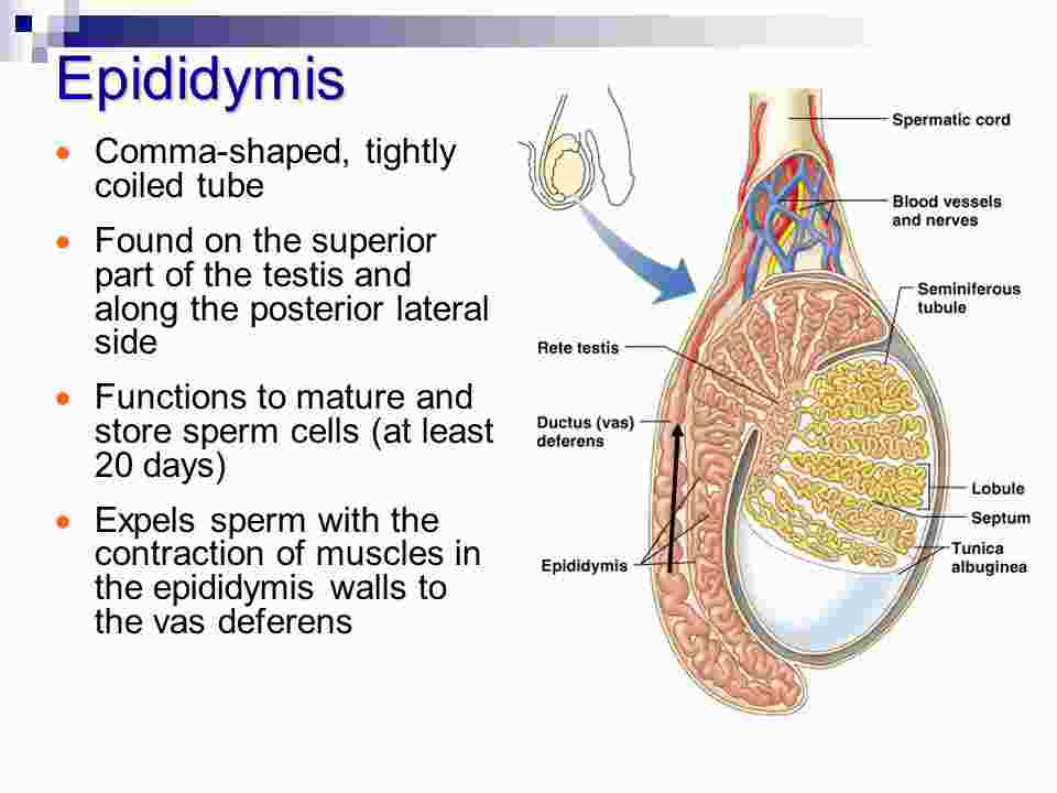
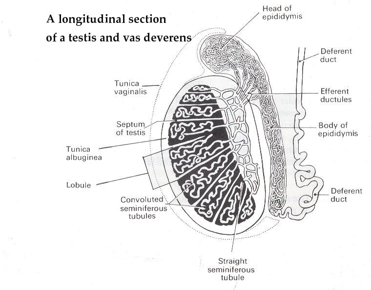
Anatomy of the Epididymis. The epididymis is 5-10 mm thick and extends from the upper to the caudal pole of the testis. The epididymis can be divided into caput, corpus, and cauda. A thin capsule and the serosa of the tunica vaginalis cover the epididymis. 8-10 efferent ducts transport the sperm from the rete testis into the epididymal duct.
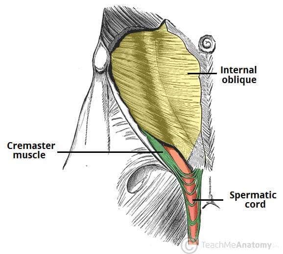
The Spermatic Cord Course Fascia Contents TeachMeAnatomy
It is the small, muscular sac that contains and protects the testicles, blood vessels, and part of the spermatic cord. The scrotum is divided internally into two compartments by a septum and each.
Spermatic cord
The spermatic cord is a tube-like structure that extends from the testicle to the abdominal cavity in males. It is responsible for carrying vital structures to and from the testicle, including blood vessels, lymphatic vessels, and the vas deferens. Structure

Anatomy Spermatic cord Diagram Quizlet
It consists of the structures that allow the testis to function normally and includes the ductus deferens, arterial and venous blood vessels, lymphatic vessels, and nerves for the testis and its covers.
Spermatic Cord Contents Quiz By
Each cord is sheathed in connective tissue and contains a network of arteries, veins, nerves, and the first section of the ductus deferens, through which sperm pass in the process of ejaculation.

What I learned today... Mnemonics... Contents of the Spermatic Cord
Chronic scrotal pain (CSP) is defined as constant or intermittent pain in the scrotum lasting for more than three months. 1 CSP does not involve only testicular pain, as there may be pain involving the epididymis, vas deferens or adjacent paratesticular structures.