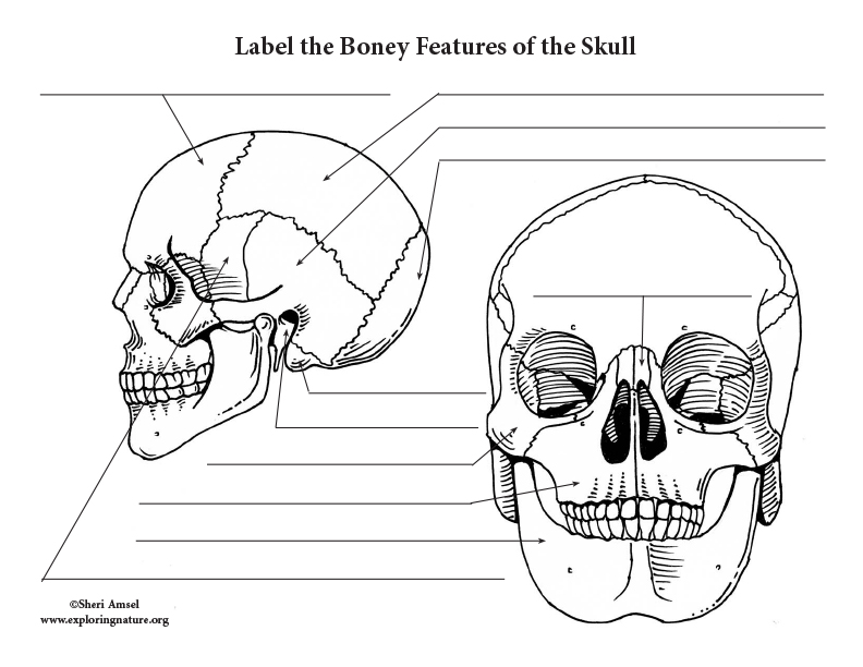
Skull Labeling
The cranium (skull) is the skeletal structure of the head that supports the face and protects the brain. It is subdivided into the facial bones and the brain case, or cranial vault ( Figure 7.3 ). The facial bones underlie the facial structures, form the nasal cavity, enclose the eyeballs, and support the teeth of the upper and lower jaws.
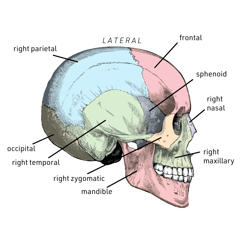
43 skull labeling worksheet answers Worksheet Master
Labeling Exercises. Skeleton-Anterior View. Skeleton-Posterior View. Lower Skeleton. Upper Skeleton-Anterior View.
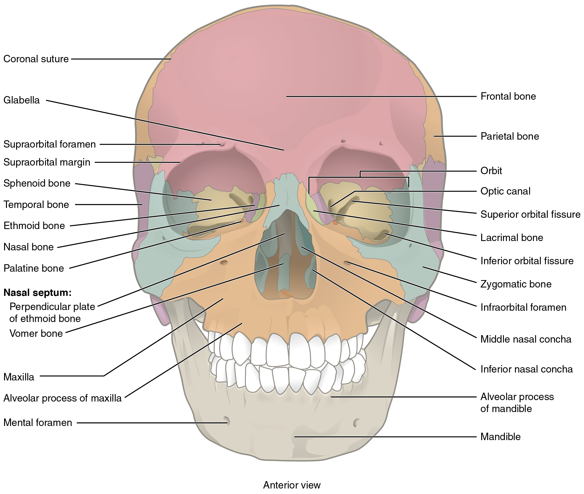
7.2 The Skull Anatomy and Physiology
1/13 Synonyms: none In this article we will be focusing on the foramina and fissures located on the inside and floor, or base, of the skull. In a nutshell, a foramen means a hole that can allow various structures to pass through them, ranging from nerves all the way to vessels.
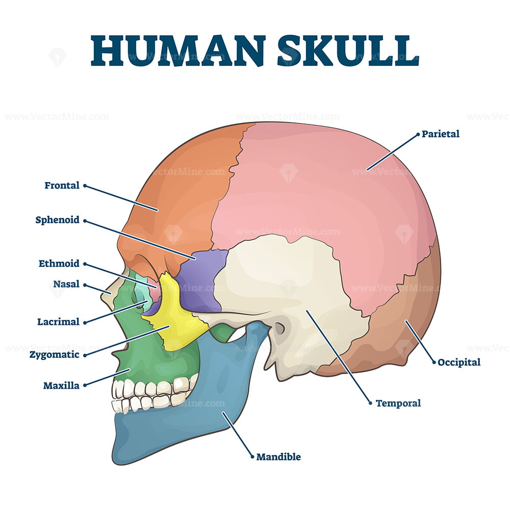
Human skull bones skeleton labeled educational scheme vector illustration VectorMine
Labeling the Adult Skull Bones by birdb08 10,250 plays 16 questions ~40 sec English 16p 12 too few (you: not rated) Tries 16 [?] Last Played February 22, 2022 - 12:00 am There is a printable worksheet available for download here so you can take the quiz with pen and paper. From the quiz author Labeling the adult skull bones - front view. Remaining

Labeled Diagrams Of Skull
Skull Label (remote) This activity was designed for anatomy and physiology with students working remotely during the 2020 pandemic. Students are given a short overview of the skull during virtual class and then encouraged to watch the Pop Up Biology video which explains the features of the skull (foramen, condyles, process…etc.)
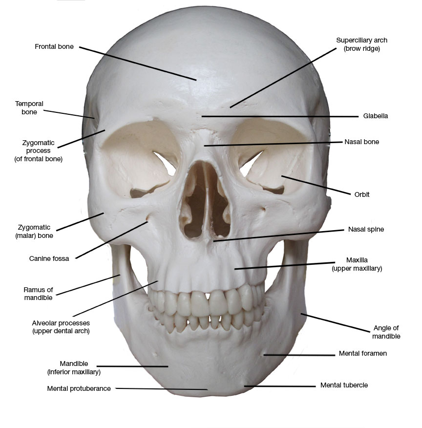
Kreated by Krause Artistic Anatomy Part 1 Frontal Skull Bones
creates a bridge-like structure that connects the temporal bone with the zygomatic bone forming part of the zygomatic arch. Above: Markings of the cranium with the following views: (A) anterior view, (B) lateral view of the left side of the skull, (C) posterior view, and (D) lateral view of the right side of the skull.
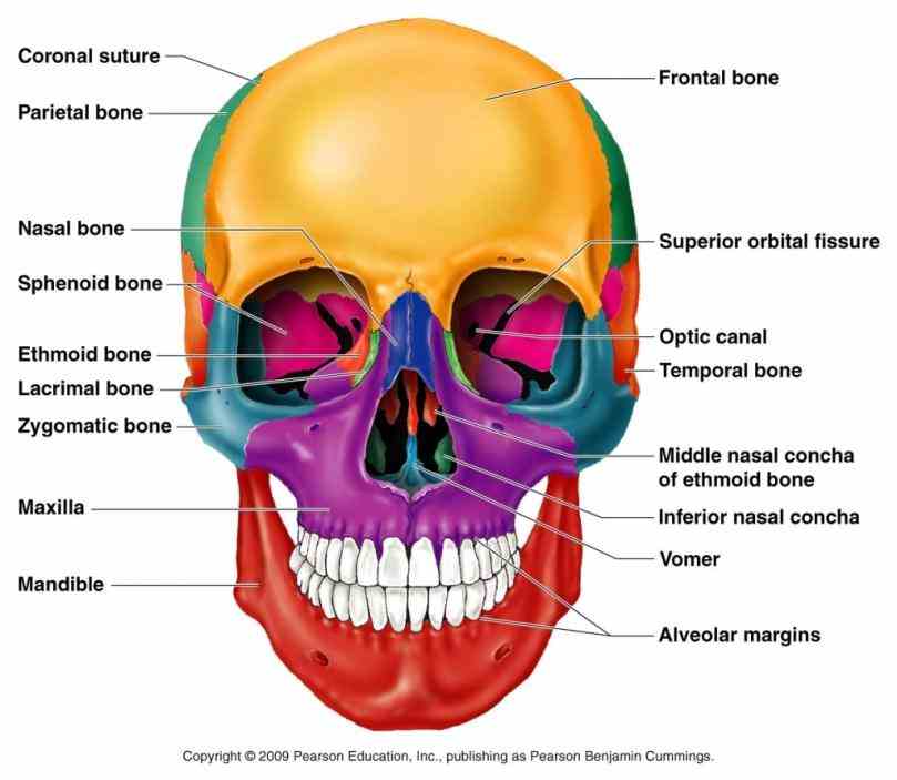
Labeled Diagrams Of Skull
1 - the skeleton : test your knowledge of the bones of the full skeleton 2 - the axial skeleton : How about the bones of the axial skeleton? 3 - the skull : Do you know the bones of the skull? 4 - the spine : Test your knowledge of the bones of the spine 5 - the hand : can you name the bones of the hand?

Medical and Health Science Anatomy of Skull
Labeled Skull Diagram. The idea behind using labeled diagrams is to get an overview of all of the structures within a given area. When it comes to testing your memory of these structures, previously having seen them altogether as a group should help you to remember them more easily. Before you use our skull diagrams free to download below, it.

Skull Diagram Labelled · Free vector graphic on Pixabay
Figure 1. Parts of the Skull. The skull consists of the rounded brain case that houses the brain and the facial bones that form the upper and lower jaws, nose, orbits, and other facial structures. Watch this video to view a rotating and exploded skull, with color-coded bones.
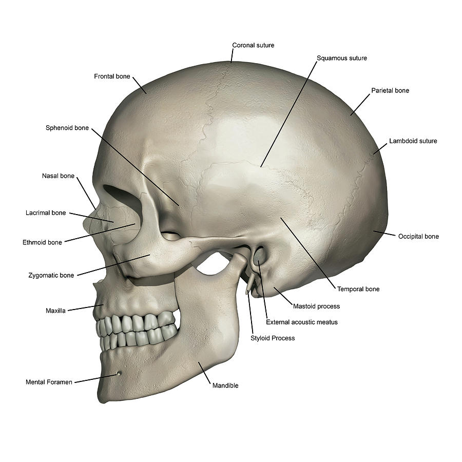
Lateral View Of Human Skull Anatomy Photograph by Alayna Guza Fine Art America
The skull is the skeletal structure of the head that supports the face and protects the brain. It is subdivided into the facial bones and the cranium, or cranial vault ( Figure 7.3.1 ). The facial bones underlie the facial structures, form the nasal cavity, enclose the eyeballs, and support the teeth of the upper and lower jaws.

Labeled Diagrams Of Skull
Official Ninja Nerd Website: https://ninjanerd.orgNinja Nerds!In this lecture Professor Zach Murphy will present on the anatomy of the skull through the use.
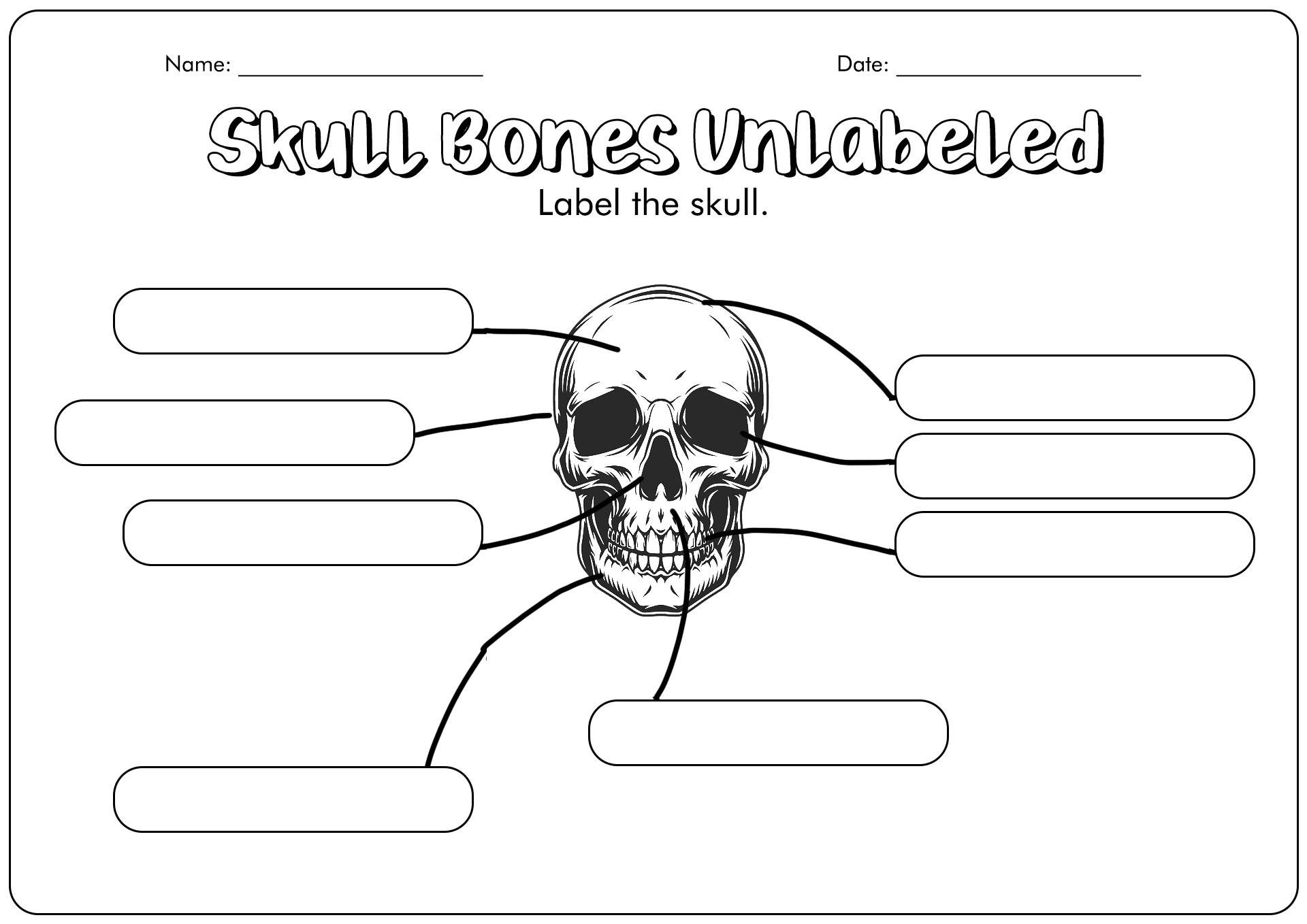
16 Skull Labeling Worksheets Free PDF at
The skull is a bony structure that supports the face and forms a protective cavity for the brain. It is comprised of many bones, which are formed by intramembranous ossification, and joined by sutures (fibrous joints).. The bones of the skull can be considered as two groups: those of the cranium (which consist of the cranial roof and cranial base) and those of the face.

Bones of the Human Skull photo Human anatomy and physiology, Anatomy bones, Basic anatomy and
Labeling the Bones of the Skull by birdb08 101,821 plays 19 questions ~50 sec English 19p More 88 3.17 (you: not rated) Tries 19 [?] Last Played January 8, 2024 - 05:57 PM There is a printable worksheet available for download here so you can take the quiz with pen and paper. From the quiz author Labeling the bones of the adult skull - side view

Printable Skull Anatomy Coloring Pages Printable World Holiday
Foramina and contents Anterior cranial fossa Middle cranial fossa Posterior cranial fossa Anterior (frontal) view Lateral (side) view Posterior view Superior view Base of the skull (inferior view) Foramina summary Sources Related articles + Show all Components and features Sagittal suture Sutura sagittalis 1/3

7.2 The Skull Douglas College Human Anatomy and Physiology I (1st ed.)
Lateral Left. Lateral Right. Skull Labeling

The Bones of the Skull Human Anatomy and Physiology Lab (BSB 141)
Pictures of skulls that are labeled for reference. Skull is pictured from many angles. Skull Labeling - Answer Key. 1. Coronal Suture 2. Frontal 3. Parietal 4. Nasal 5. Squamosal Suture 6. Ethmoid 7. Lacrimal 8. Sphenoid 9. Lamdoidal Suture 10. Occipital 11. Temporal 12. Zygomatic 13. Maxilla 14. Mandible